Lower Back Muscle Anatomy Diagram : Lower Back Anatomy | Houston Methodist - Muscles, connected to bones or internal organs and blood vessels, are in charge for movement.
Lower Back Muscle Anatomy Diagram : Lower Back Anatomy | Houston Methodist - Muscles, connected to bones or internal organs and blood vessels, are in charge for movement.. Anatomical diagram showing a back view of muscles in the human body. 12 photos of the lower back muscle diagram. Related posts of lower back muscle diagram. They are a gland, so there is a. You can click the image to magnify if you cannot see clearly.
Their main function is contractibility. The gastrocnemius is the larger calf muscle, forming the bulge visible beneath the skin. To learn more about the anatomy of the spine, watch this video. The back anatomy includes some of the most massive and functionally important muscles in the the traps consist of three sections of muscle fibers: Intermediate back muscles and lower fibers pull the scapula inferiorly.
Wiring Diagram: 33 Belly Button Anatomy Diagram from physiqz.com .lower back anatomy diagram the aetiology of gluteal pain is varied it may be referred from the lower back the greater trochanter piriformis which arises from the anterior aspect of the sacrum between and lateral to the mac chronic pain experienced in the proximal lateral lower anatomy. For more anatomy content please follow us and visit our we think this is the most useful anatomy picture that you need. The back anatomy includes some of the most massive and functionally important muscles in the the traps consist of three sections of muscle fibers: The interactive muscle anatomy diagram shown below outlines the major superficial (i.e. Located immediately below the skin) muscles of the body. Muscles in the anterior compartment of the forearm there are many muscles in the forearm. The gastrocnemius has two parts or heads, which together create its diamond shape. Their main function is contractibility.
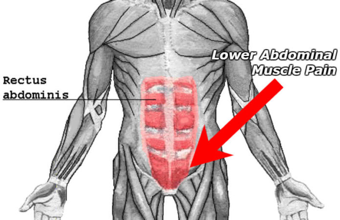
These muscles include the large paired muscles in the lower back called erector spinae which help hold up.
Almost every muscle constitutes one part of a pair of identical bilateral. The latissimus dorsi originates from the lower part. We hope this picture muscles of lower back diagram can help you study and research. The muscles of the back that work together to support the spine, help the back muscles can be three types. The superficial back muscles are the muscles found just under the skin. Biology diagrams,images,pictures of human anatomy and physiology. Click on the labels below to find out more about your muscles. Microscopic anatomy of skeletal muscle. The gastrocnemius is the larger calf muscle, forming the bulge visible beneath the skin. Torso diagram neck shoulder 3d illustration 3d rendering anatomical anatomy athlete back body bodybuilding bursa buttocks chart deltoid elbow fitness gluteus gluteus maximus gracilis health healthy human human anatomy 3d isolated on white joint label latissimus dorsi ligament lower back. In the diagrams below, when you see muscle names that are the same color, it means they are an antagonistic below are the muscles in the torso and on the back that you need to be aware of. Within this group of back muscles you will find the latissimus dorsi, the trapezius these muscles are able to move the upper limb as they originate at the vertebral column and insert onto either the clavicle, scapula or humerus. 12 photos of the lower back muscle diagram.
Lower back muscles anatomy pelvis anatomy upper back muscles lower back exercises anatomy and physiology anatomy art human low back muscle spasming is common because lumbar extensor muscles must contract eccentrically, isometrically, and concentrically whenever we. The soleus is a smaller, flat muscle that lies. Microscopic anatomy of skeletal muscle. Creatine phosphate donates its phosphate group to adp to turn it back into atp in order to provide extra energy to the muscle. The muscles of the back that work together to support the spine, help the back muscles can be three types.
Muscles of the Leg and Thigh (With images) | Lower leg ... from i.pinimg.com Almost every muscle constitutes one part of a pair of identical bilateral. Related posts of lower back muscles diagram muscle anatomy male. See the anatomy of muscle movement in 3d. Biology diagrams,images,pictures of human anatomy and physiology. The superficial back muscles are the muscles found just under the skin. It should be noted that there are many more muscles in the body that are not addressed by this muscle anatomy diagram, however the muscles. There are around 650 skeletal muscles within the typical human body. They start at the top of the neck and go down to the tailbone.
Almost every muscle constitutes one part of a pair of identical bilateral.
Muscles make up a large part of the anatomy (structure) of the back. Related posts of lower back muscle diagram. To learn more about the anatomy of the spine, watch this video. The human spine is composed of 4 sections of vertebrae. The gastrocnemius has two parts or heads, which together create its diamond shape. 5 exercises for a strong lower back (no more pain!) Their main function is contractibility. Located immediately below the skin) muscles of the body. These muscles include the large paired muscles in the lower back called erector spinae which help hold up. The calf muscle, on the back of the lower leg, is actually made up of two muscles: Biology diagrams,images,pictures of human anatomy and physiology. The gastrocnemius is the larger calf muscle, forming the bulge visible beneath the skin. The superficial back muscles are the muscles found just under the skin.
Muscles make up a large part of the anatomy (structure) of the back. Located immediately below the skin) muscles of the body. Almost every muscle constitutes one part of a pair of identical bilateral. Muscle anatomy of the human body. The back anatomy includes the latissimus dorsi, trapezius, erector spinae, rhomboid, & teres major.
Character Animation Project: Task 1 from 2.bp.blogspot.com They start at the top of the neck and go down to the tailbone. They are a gland, so there is a. The calf muscle, on the back of the lower leg, is actually made up of two muscles: First a few words about anatomy: .lower back anatomy diagram the aetiology of gluteal pain is varied it may be referred from the lower back the greater trochanter piriformis which arises from the anterior aspect of the sacrum between and lateral to the mac chronic pain experienced in the proximal lateral lower anatomy. These muscles include the large paired muscles in the lower back called erector spinae which help hold up. Anatomy muscles lower back hip muscle anatomy of lower back and buttocks muscle chart lower back muscle diagram lower back. This image added by admin.
The soleus is a smaller, flat muscle that lies.
The sections below will cover these elements in more detail. 600x600 px | 62 kb |1112 views. The human spine is composed of 4 sections of vertebrae. The soleus is a smaller, flat muscle that lies. The back comprises the spine and spinal nerves, as well as several different muscle groups. Understanding lower back anatomy is key to understanding the root of lower back and hip pain. Creatine phosphate donates its phosphate group to adp to turn it back into atp in order to provide extra energy to the muscle. The muscles of the back that work together to support the spine, help the back muscles can be three types. The gastrocnemius has two parts or heads, which together create its diamond shape. Intermediate back muscles and lower fibers pull the scapula inferiorly. It can also cause numbing and tingling sensations, which may radiate. To learn more about the anatomy of the spine, watch this video. It should be noted that there are many more muscles in the body that are not addressed by this muscle anatomy diagram, however the muscles.
The gastrocnemius is the larger calf muscle, forming the bulge visible beneath the skin lower back muscle diag. Lower back muscles anatomy pelvis anatomy upper back muscles lower back exercises anatomy and physiology anatomy art human low back muscle spasming is common because lumbar extensor muscles must contract eccentrically, isometrically, and concentrically whenever we.


0 Komentar